What Wildlife Workers Should Know About Avian Anatomy

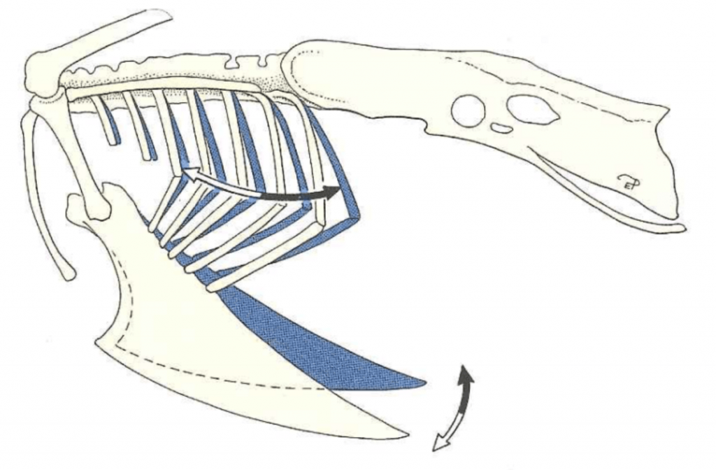
Figure 1: Motion of the keel and ribs during inspiration (white arrows) and expiration (black arrows). Credit: König et al.

As veterinary students, virtually all the anatomy that we learn in the core curriculum is mammalian anatomy, but most of the patients coming into the Janet Swanson Wildlife Health Center are birds.   To help fellow veterinary students and other wildlife workers, I have created this post as a brief guide to avian anatomy, focusing specifically on aspects which are directly relevant to providing routine avian care at a wildlife hospital.


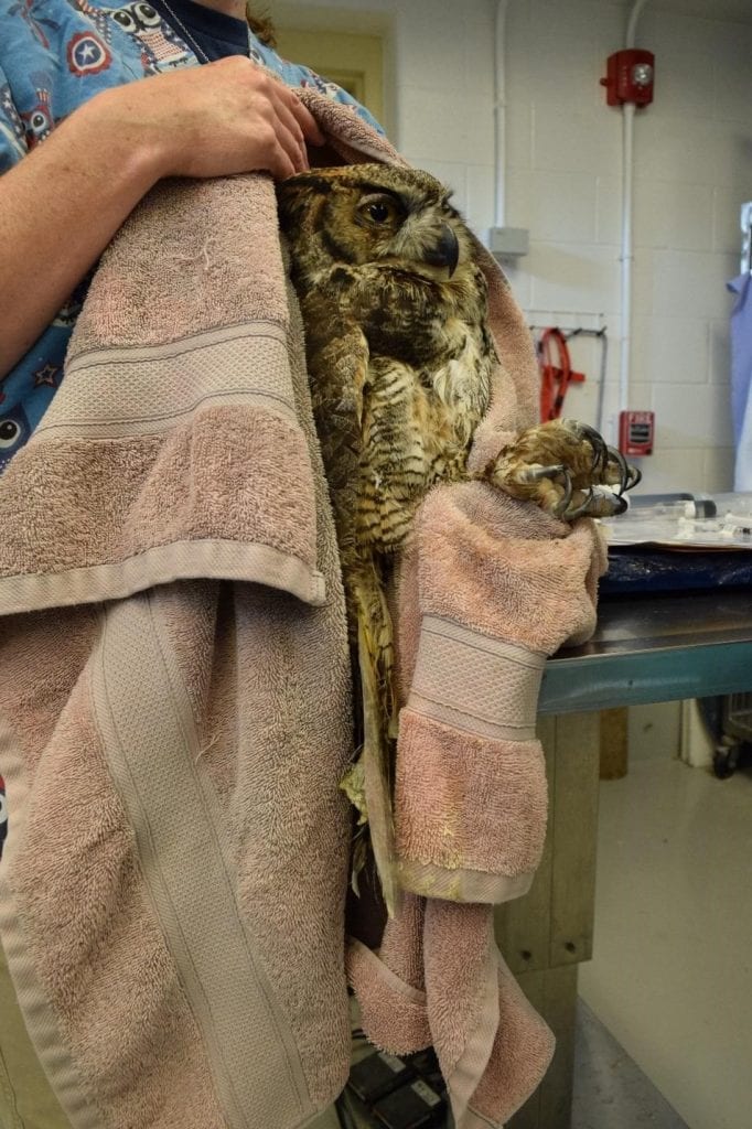
Figure 3: Example of a good hold for a raptor. Notice how, like the blue jay, this great horned owl is restrained from the back and by the feet. Credit: Jonathan Gorman

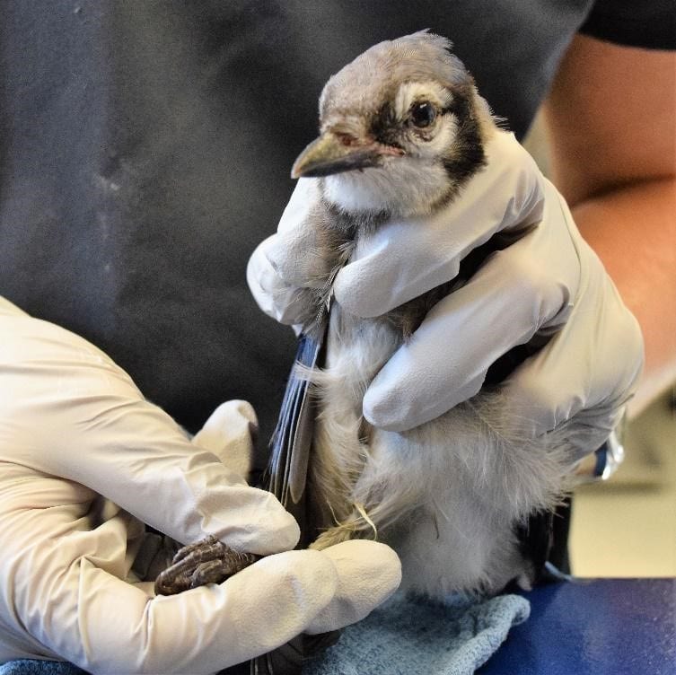
Figure 2: Example of a good hold for a passerine. Notice that this blue jay is primarily restrained from the back and by the legs so as not to put pressure on the sternum. Credit: Jonathan Gorman

Respiratory Anatomy


In birds, the sternum exists as a large fused structure called the keel.  Unlike mammals, birds do not have a diaphragm; during inspiration they rely on the external intercostal and levator costarum muscles which move the ribs laterally and the sternum cranio-ventrally (Figure 1).  When handling birds, it is very important not to put pressure on the keel since this prevents these movements and can potentially cause asphyxiation.  It is therefore necessary to use appropriate restraining methods that don’t place pressure on the keel (Figures 2 and 3).  During inspiration, the weight of the viscera helps to pull the sternum in the cranio-ventral direction.  However, when a bird is flipped on their back, the viscera weighs the sternum down making inspiration more difficult.  For this reason, extra caution must be taken when placing birds on their backs for long periods of time.

In the mammalian respiratory system, air enters the lungs during inspiration and exits the lungs during expiration in a bidirectional manner .  Birds instead have a unidirectional system of air flow that relies on thin-walled cavities attached to the lungs called air sacs. This allows fresh air to pass through the lungs during both inspiration and expiration (Figure 4).  The air sacs extend throughout most of a bird’s body cavity (Figure 5), covering the viscera, and even projecting into major bones.  This is important to keep in mind during subcutaneous injections, so that one does not inject into an air sac.  Pulling back on the plunger to check for negative pressure before giving a subcutaneous injection will ensure that an air sac has not been penetrated.

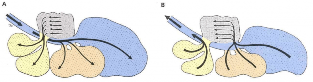
Figure 4: Schematic of avian respiration. (A) During inspiration, air in the trachea flows to the caudal air sacs (caudal thoracic and abdominal air sacs) while air in the lungs flows to the cranial air sacs (cranial thoracic and clavicular air sacs). (B) During expiration, the air in the caudal air sacs flows through the lungs while the air in the cranial airs sacs flows out the trachea. Credit: König et al.

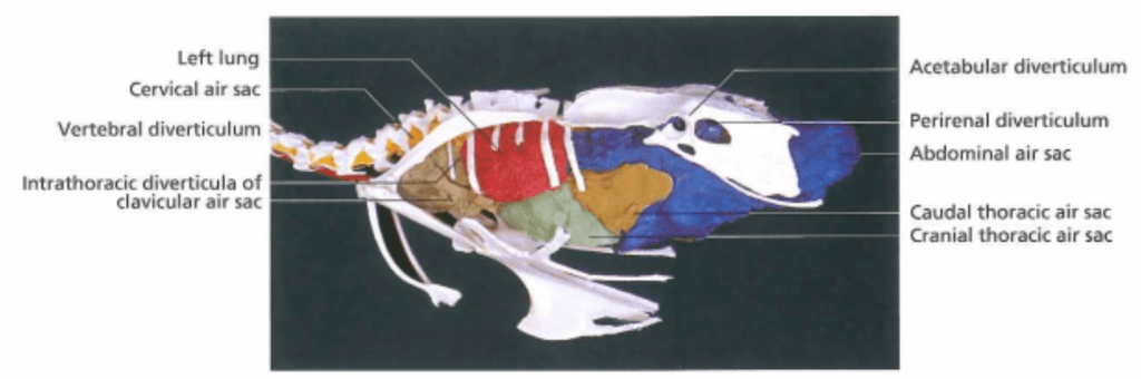
Figure 5: Visual representation of the air sacs in a chicken. Credit: König et al.


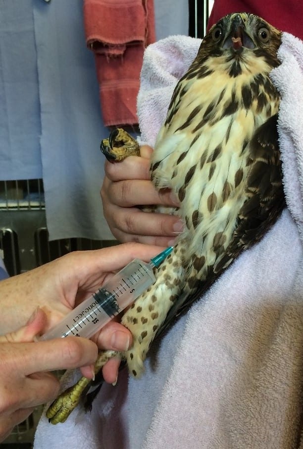
Figure 6: Subcutaneous injection into the inguinal fold. Courtesy of Dr. Childs-Sanford

Subcutaneous Injections

For birds, there are two main sites of subcutaneous injection used at the Wildlife Health Center.  The preferred site for subcutaneous fluid administration is the inguinal fold, which is located at the proximal aspect of the femur (Figure 6).  Another site used for injections in large birds is the interscapular region, which is over the back and between the shoulders.  If injecting here, it is important not to get too close to the neck, since this would risk puncturing the cervical air sac.


Figure 7: Anatomy of the avian oral cavity. Credit: König et al.

Gastrointestinal Anatomy

When giving food or medications to a bird by gavage  (i.e. using a tube or crop needle that goes down the throat), it is very important to place the instrument down the esophagus and not the trachea.  The esophagus in birds has a very broad opening at the back of the mouth.  In contrast, the glottis (i.e. tracheal opening) appears as a slit on the floor of the mouth just caudal to the tongue (Figure 7).  In order to avoid aspiration of fluid when giving liquid medication, the medicine should be administered where it will not enter the glottis, either behind the glottis or at the tip of the beak where it will flow to the sides of the mouth.

Unlike in mammals, the avian esophagus runs down the right side of the neck.  When inserting a tube or crop needle, correct instrument placement can be verified by palpating the instrument through the esophagus to make sure it is not in the trachea – if it is in the correct place, one should feel two tubes: the gavage tube or crop needle in the esophagus, and the trachea.  If only one tube can be felt, the instrument may be inside the trachea.  Additionally, if the instrument is placed correctly one should be able to see that the opening of the glottis is clear (Figure 8).

Some species of birds have an enlargement of the esophagus at the level of the thoracic inlet called a crop.  The crop temporarily stores ingesta before it enters the proventriculus (i.e. the glandular stomach).  When using a tube or crop needle to gavage a bird, the tip of the instrument should be all the way in the crop to avoid aspiration.  It is therefore a good idea to take note of how much length is needed to reach the thoracic inlet so that the instrument is placed far enough down the esophagus.

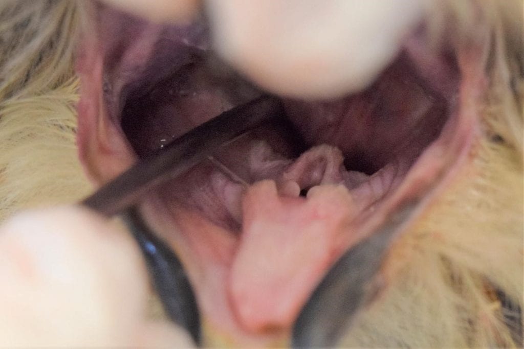
Figure 8: Correct placement of a crop needle for a great horned owl. The needle is entering the esophagus and the glottis is clear. Credit: Jonathan Gorman


Anatomy of Elimination

The final part of the gastrointestinal tract in birds, the cloaca, is divided into three parts: the coprodeum, the urodeum, and the proctodeum (Figure 9).  Gastrointestinal contents enter the coprodeum from the rectum.  They then pass through the urodeum where the urinary and reproductive tracts open.  The proctodeum is the final segment of the cloaca, and it houses the copulatory organ (phallus) in males, something important to note when sexing some species of birds.

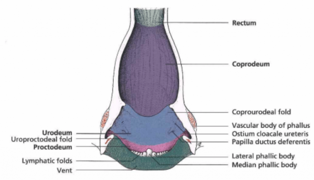
Figure 9: Anatomy of the avian cloaca. Credit: König et al.

Because the ureters open into the cloaca, birds pass their urine with their feces; this is important to note when evaluating feces and urates.  For instance, a bird with excessively watery excreta may have diarrhea or it may have polyuria.  Diarrhea would present as liquid feces while polyuria would present as an excess of liquid surrounding a relatively firm fecal mass.  One must take into account that the normal appearance of feces and urates varies significantly by species and diet (Figure 10).

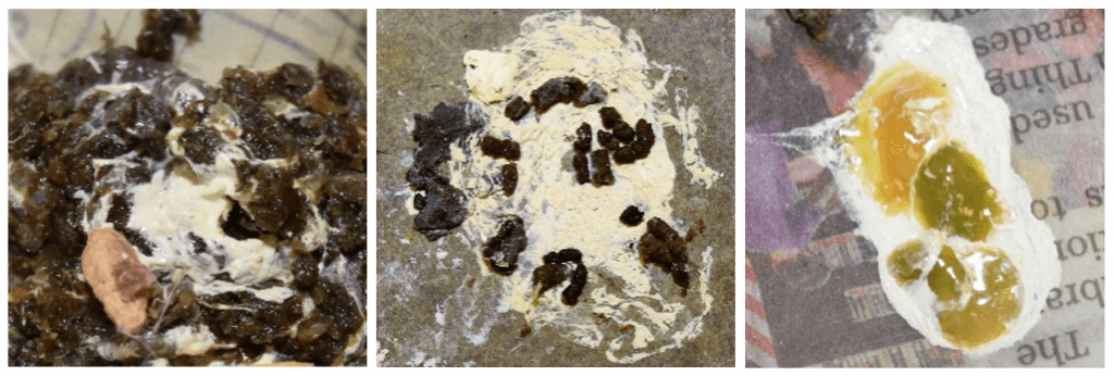
Figure 10: Different images of ‘normal’ feces and urates from different types of birds. The one on the left from a pigeon is typical of granivores; the feces is firm and easily distinguished from the urates which are thick and pasty. The middle image from a great horned owl is typical of raptors which have thick or thin viscous feces mixed with urine. ‘Soft-feeders’, namely insectivores and frugivores, produce feces and urates like that presented on the right from a blue jay. The feces is thin but the color and consistency can vary greatly depending on diet. Credit: Jonathan Gorman


Integument System

The beak is covered in a hard keratinized  sheath, the rhamphotheca.  Most birds have an aggregation of sensory receptors at the tip of the beak known as the bill tip organ.  This organ is especially well-developed in ducks and geese but is absent in pigeons and sparrows.  It is important to note when working around the beak that this can be a sensitive area.

While birds do not have sweat glands, some of them do have an important gland located above the tail called the uropygial gland.  The uropygial gland produces an oily secretion which certain birds, particularly waterfowl, spread around over their feathers during preening to create a waterproof film.  This waterproof film can be degraded by urates, which is why it is important to keep housing for waterfowl clean.  This is especially important for birds that can no longer preen themselves due to injury.


Acknowledgements

I would like to thank Dr. Childs-Sanford for providing feedback and advice on this post.   I would also like to thank Isabel Jimenez (’19) for helping me come up with ideas and for sharing her knowledge on wildlife care.  Lastly, thank you to the Janet L. Swanson Wildlife Health Center, its staff, and its volunteers, for providing me with the inspiration and photo opportunities for this post.


References

König, Horst E, Rüdiger Korbel, Hans-Georg Liebich, Hermann Bragulla, Klaus-Dieter Budras, Romay A. Carretero, Gerhard Forstenpointner, Corinna Klupiec, Johann Maierl, Maren Meiners, Ivan Míšek, Christoph K. W. Mülling, Beltrán M. Navarro, Alexander Probst, Sven Reese, Jesus Ruberte, Ingrid Walter, Gerald Weissengruber, and Grammatia Zengerling. Avian Anatomy: Textbook and Colour Atlas. Sheffield: 5M Publishing, 2016. Print.


Wildlife Health comes to New York City – Investigating Lead Levels in Pigeons

Credit: Jennifer Morrow, https://www.flickr.com/


Wildlife in New York City. It sounds like an oxymoron, but every year, concerned New Yorkers find thousands of injured animals lying on the sidewalk. They take to Google and end up at the only wildlife rehabilitation center in NYC – the Wild Bird Fund. I was one of those people.

In 2009, two children brought a sparrow into the dog & cat clinic that I worked at. The bird wasn’t putting any weight on its right leg. After my shift was over, I made the two-hour subway ride to the Upper West Side and stepped through the front door of the Wild Bird Fund. Hens roamed freely in the lobby. A gull honked from its perch on the chair next to me. Mourning doves cooed from huge window aviaries, and a rehab worker bustled by cradling a swan, one hand supporting its long neck. I was instantly hooked. The very next week, I attended orientation to become a volunteer.

Although the Wild Bird Fund accepts all animals, more than half of its patients belong to just one species. It’s the most commonly sighted animal in New York City – the infamous pigeon, Columba livia, or, “rat with wings”.  There are over a million in NYC alone. That means thousands of sick pigeons arrive at the Wild Bird Fund every year. After volunteering for a while, I became interested in learning more about lead poisoning, one of the most common ailments among our patients.

Map of pigeon mean blood lead levels in Manhattan by zip code.

For my undergraduate senior thesis project, I decided to compile all of the blood lead levels for pigeons measured at the Wild Bird Fund from 2011 onwards. Then, I mapped these results across different neighborhoods in NYC. Sorting through and geolocating thousands of hand-written records took me the better part of an entire summer. Turns out it wasn’t in vain – I found consistently high levels of lead poisoning in small pockets of the city, areas such as Lower Manhattan and the Gowanus Canal in Brooklyn. My results also matched child lead poisoning reports published by the US Department of Health in 2010 and 2014. Sick children and sick pigeons live in the same neighborhoods.

It’s still unclear where this lead is coming from, although it’s most likely a combination of several different sources, including leaded paint, leaded gasoline, and small airplane fuel. The Wild Bird Fund data also revealed another trend: lead poisoning is significantly higher in the summer. This correlates with lead poisoning studies done on human children; Laidlaw et al. (2005) suggest that soil humidity is lower in the summer, leading to increased suspension of and exposure to lead dust.

In the end, it’s hard to look past some of the similarities between human and animal pathology, especially when we live in the same neighborhoods, breathe the same air, and eat the same food. (If you’ve never seen a pigeon chowing down on a discarded pretzel, have you truly seen New York City?) Mine is just one of many studies being published every year about human-ecosphere interaction. One emerging field involves the use of animals as bioindicators for human health. The bioindicator is the proverbial canary in the coal mine; if the wildlife falls ill, we may expect humans to fall ill as well.

Just like any other time science happens, I submitted my paper with more questions than I had answers. Wildlife is suffering, and it is by no uncertain terms our doing. It’s hard not to notice this when three of the most common problems treated at the Wild Bird Fund are lead poisoning, collision with windows, and cat attacks. Shortly after my study was published in Chemosphere, it was picked up by The New York Times and a slew of other online papers. In particular, The New York Times debated the true efficacy of predicting human disease using pigeon lives. As I read the news article, my heart sank.

I’d written a paper on pigeon health, but I was getting the impression that very few people cared if it didn’t have immediate applications to human medicine. It wasn’t entirely their fault — I had, after all, dedicated many pages of my study to drawing comparisons between pigeon and human child lead toxicity. But as accurate as these comparisons were, I’d done them in part because I hadn’t been confident that anyone would notice a study on just pigeon lead poisoning. I thought my results were important, and I wanted them to be published; writing about human medicine would help that process.

It’s not that I don’t care about human medicine — far from it. We should raise our voices, loudly and clearly, when a human is poisoned. And we should raise our voices, loudly and clearly, when an animal is poisoned as well. Lead toxicity or otherwise, it’s becoming more and more obvious that connections between human and animal health exist everywhere we turn. I guess this is a call to everyone to care just a bit more about what happens to the life that we’re surrounded by: the earthworms tunneling underfoot, the warblers migrating south, and yes, maybe that pigeon strutting past too, even if it just pooped on your windshield.


The paper published from Fayme’s research can be found here.  Click here to read the New York Times article about it.


ABOUT THE AUTHOR:

Fayme Cai, class of 2022, was born and raised in NYC and graduated from Columbia University with a Bachelor’s in Ecology & Evolution and a minor in Psychology. Although it’s still up in the air for now, she’s mostly interested in small animal and companion exotic animal medicine.

Six speakers discuss different aspects of conservation medicine at the first annual Wildlife Health Day

Dr. Elizabeth Buckles discussing the histopathology of white-nose syndrome, a fungal disease affecting North American bats.

Zack Dvornicky-Raymond DVM (’19)

On Saturday, six speakers shared their experiences in conservation, wildlife, and One Health. The topics were wide-ranging, covering conservation of endangered species, zoo animal nutrition, plastic waste, and honeybee health. For every topic, the speakers highlighted the importance of wildlife health and the role of the veterinarian.

Zack Dvornicky-Raymond (’19), kicked off Wildlife Health Day with a talk that drew from both his personal experience and his knowledge of the conservation field. Zack first described his Expanding Horizons experience in Namibia where he used his veterinary skills to help the Cheetah Conservation Fund with their guard dog breeding program. He then discussed Assisted Reproductive Technologies (ARTs) and their use in breeding endangered species, drawing from his experience studying canine reproduction at the Travis Lab. Zack also drew from his experience at the Smithsonian, where he investigated better ways to transport sperm for the endangered Przewalski’s horse, information critical for preserving the genetic diversity of the species. Zack then discussed the role of contraception-based wildlife management, before finishing off with a reflection about the impact of human population growth on the state of the natural environment.

Dr. Sara Childs-Sanford discussed the unique challenges of managing nutrition in captive wildlife. Many of the nutritional problems that zoo animals face stem from our lack of knowledge of the specific nutritional requirements for these understudied species. Dr. Childs-Sanford did leave the audience with some solutions, citing successful attempts in nutrition research: improving the reproductive success of maned wolves and helping pangolins survive in captivity.

Mariah Beck (’20)

Drawing on her research and coursework, Mariah Beck (’20) shifted the focus to environmental health with her talk on our use and waste of plastic, and its effect on ocean life. She cited three main ways that plastic waste harms marine animals: entanglement, ingestion, and toxicity. Not only do plastics harm iconic species like whales and turtles, but their toxicity also harms animals like mussels which provide important ecosystem services. Mariah called on veterinarians to advocate for reducing the amount of plastic waste in the ocean and pitched ideas to help achieve this goal.

Dr. Elizabeth Buckles dove into a case study on white-nose syndrome in bats, drawing from her own experience as a veterinary pathologist who helped identify the cause of the outbreak. This inspired several principles for working with understudied species including “know your species,” “reach out to experts,” and “be creative.” Dr. Buckles finished with several wildlife pathology anecdotes, including one about how the CDC ignored warnings from veterinarians about the arrival of West Nile Virus to the United States. These examples illustrated the need for veterinarians to speak up to protect both human and wildlife health.

The conversation moved to honeybee health when Dr. Robin Radcliffe gave a lecture on colony collapse and the light that wild honeybee colonies can shed on it. Curiously, wild honeybee populations have not suffered from all of the problems that face captive bee colonies. For instance, as Dr. Radcliffe explained, wild honeybees have developed behavioral immunity to the Varroa mite which has devastated captive bee populations. His main  message was that there is a need for greater monitoring of colony health by the veterinary community, something the Cornell College of Veterinary Medicine is facilitating by adding a new distribution course on apiary medicine.

Dr. Sharon Deem speaks at Wildlife Health Day 2018.

Dr. Sharon Deem, wildlife veterinarian for the St. Louis Zoo, finished off the day as the keynote speaker. She rehashed many of the issues brought up by other speakers, while also sharing her perspective on the most important issues in wildlife health. Dr. Deem emphasized One Health, drawing on her research on diseases of camels being used as livestock in Kenya.  She left the audience with advice for future wildlife veterinarians.

Wildlife Health day drew dozens of vet students and faculty, giving members of the Cornell Veterinary community a chance to learn about the opportunities and challenges within the field. These types of events aim to increase levels of awareness and emphasize the importance of veterinarians in conservation and One Health.

EVENT: WILDLIFE HEALTH DAY (time and location updated)

During this 1st annual event, ZAWS will be bringing a selection of diverse speakers, lecturing on topics ranging from the role of reproduction in conservation, the effects of plastic on biodiversity loss, and the importance of nutrition for wildlife conservation.

Featuring Keynote Speaker: Dr. Sharon Deem, Director of the Saint Louis Zoo Institute for Conservation Medicine

Veterinary Medicine in the Anthropocene Epoch:

The lecture will focus on the current challenges of the 21st century: minimizing the loss of biodiversity, feeding 7.6 billion people without causing too much harm to the planet, and mitigating the negative impacts of climate change on animal health. The lecture will be from the perspective of a wildlife veterinarian and her 20+ years of working on free-living wildlife health issues and with zoo collection animals at AZA accredited zoos. Dr. Deem will share stories from her work with elephants in Asia and Africa, turtle species from all over the world, and disease issues at the livestock–wildlife interface. Dr. Deem will also showcase what a veterinarian starting out can do for One Health.


Other speakers include:

Mariah Beck
Jason Sifkarovski
Zack Dvornicky-Raymond
Dr. Sara Childs-Sanford
Dr. Elizabeth Buckles
Dr. Robin Radcliffe

This lecture series will be followed by dinner and the keynote presentation.

Date and Time: Saturday, February 10, 2018, 12:30pm to 8pm

Location: Cornell College of Veterinary Medicine Atrium

Register here or through the email sent out on the ZAWS listserv.


Wildlife Health: new faculty to focus on Planetary Health at Cornell CVM

Image from Dr. Osofsky’s “Explaining Planetary Health” article, One-Health Cornell Blog.

The CUCVM student community would like to extend a warm welcome to a number of phenomenal new hires in the wildlife health realm. Recently, the CVM has brought-on seven faculty and staff, with the goal of growing Wildlife Health / One Health / Planetary Health programs at a critical time in the College’s strategic planning.  The group strives to develop and apply science-based, multidisciplinary approaches to conservation, including through a focus on Planetary Health. In short, Planetary Health is a field focused on improving our understanding and applying appropriate metrics regarding the public health impacts of anthropogenic environmental change, so as to be able to inform decision-making in the land-use planning, environmental conservation, and public health policy realms. Planetary Health also provides a lens for the new CVM-led Master of Public Health program, with its first class starting in September of 2017. There have already been numerous excellent discussions and many new initiatives are underway, not only within the CVM, but throughout the University. Hopefully, new collaborative efforts will arise and continue to foster future discussions and cross-disciplinary action!

We look forward to the incredible things that will come from these new appointments, not only for Wildlife Health and Environmental Conservation at our University, but for conservation initiatives worldwide.

Please join me in welcoming:

Dr. Steven A. Osofsky, Jay Hyman Professor of Wildlife Health & Health Policy

Dr. Martin Gilbert, Senior Research Associate in Wildlife Health

Ms. Helen Lee, Wildlife Health & Health Policy – planning and operations

Ms. Shirley Atkinson, Wildlife Health & Health Policy – Southern Africa, AHEAD Program

Dr. Montira Pongsiri, Planetary Health Alliance Science Policy Advisor

Dr. María Forzán, Senior Research Associate in Wildlife Pathology

Dr. Mani Lejeune, Director of Clinical Parasitology and Senior Extension Associate at the Animal Health Diagnostic Center


ABOUT THE AUTHOR: Zack Dvornicky-Raymond is a current second-year vet student interested in wildlife conservation and One Health, and hopes to pursue a career focused on reproduction and population management in endangered/threatened wild species.