
 During the summer leading up to my third year of veterinary school, I worked with the Jane Goodall Institute (JGI) in the Republic of Congo. As part of the college’s commitment to wildlife health and international medicine, Cornell has established a partnership with JGI through the Engaged Cornell Program. Engaged Cornell gives veterinary and undergraduate students the opportunity to apply concepts learned in the classroom to field sites in developing nations across the globe. Students can elect to take an on-campus course during the spring term and apply for the opportunity to continue their studies abroad during the summer. Upon return, they take a follow-up course in the style of a seminar series, to share their work with faculty and peers, and to learn about their classmates’ experiences. Thanks to Engaged Cornell, I was able to work at Africa’s largest chimpanzee sanctuary alongside one of the world’s leading experts in the field, Dr. Rebeca Atencia.
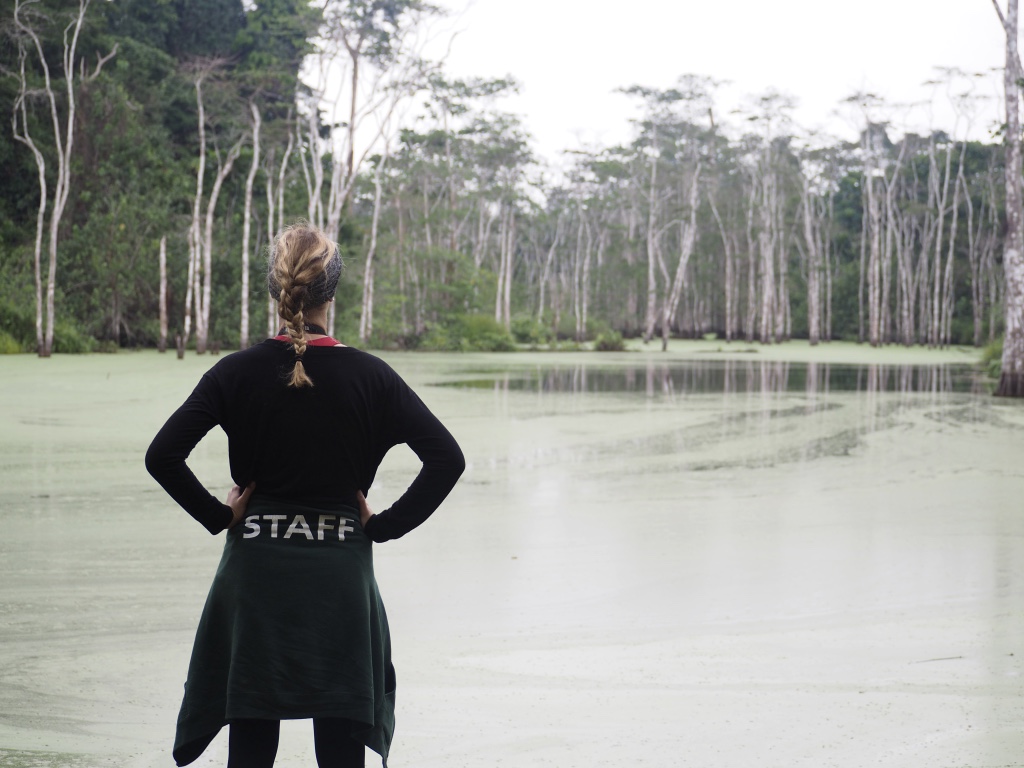
 Tchimpounga Chimpanzee Rehabilitation Center is currently home to over 130 chimpanzees, most of which fell victim to the illegal bush meat and pet trades early on in life. Through the efforts of Dr. Atencia and her team of nurses and caregivers, these animals are being given a second chance. JGI is currently working to prepare a number of these chimpanzees for release back into the forest, where they will have the opportunity to live as wild apes once again. Before that can happen, however, they need to be both physically and psychologically fit to survive the harsh realities of life in the rainforest. My efforts this summer were put towards ensuring the capability of these chimps to thrive outside of the confines of the sanctuary.
Some of my time in Congo was spent performing routine “health checks,” or comprehensive physical exams, on animals under the care of JGI. I participated in the anesthesia, general examination, cardiac evaluation, and abdominal ultrasonography of over thirty chimpanzees. I learned basic skills such as taking blood pressure measurements, giving injections, and drawing blood, and more advanced skills such as abdominal ultrasound, echocardiography, and designing anesthesia regimes. I gained invaluable hands-on veterinary experience that I truly could not have gotten anywhere else.
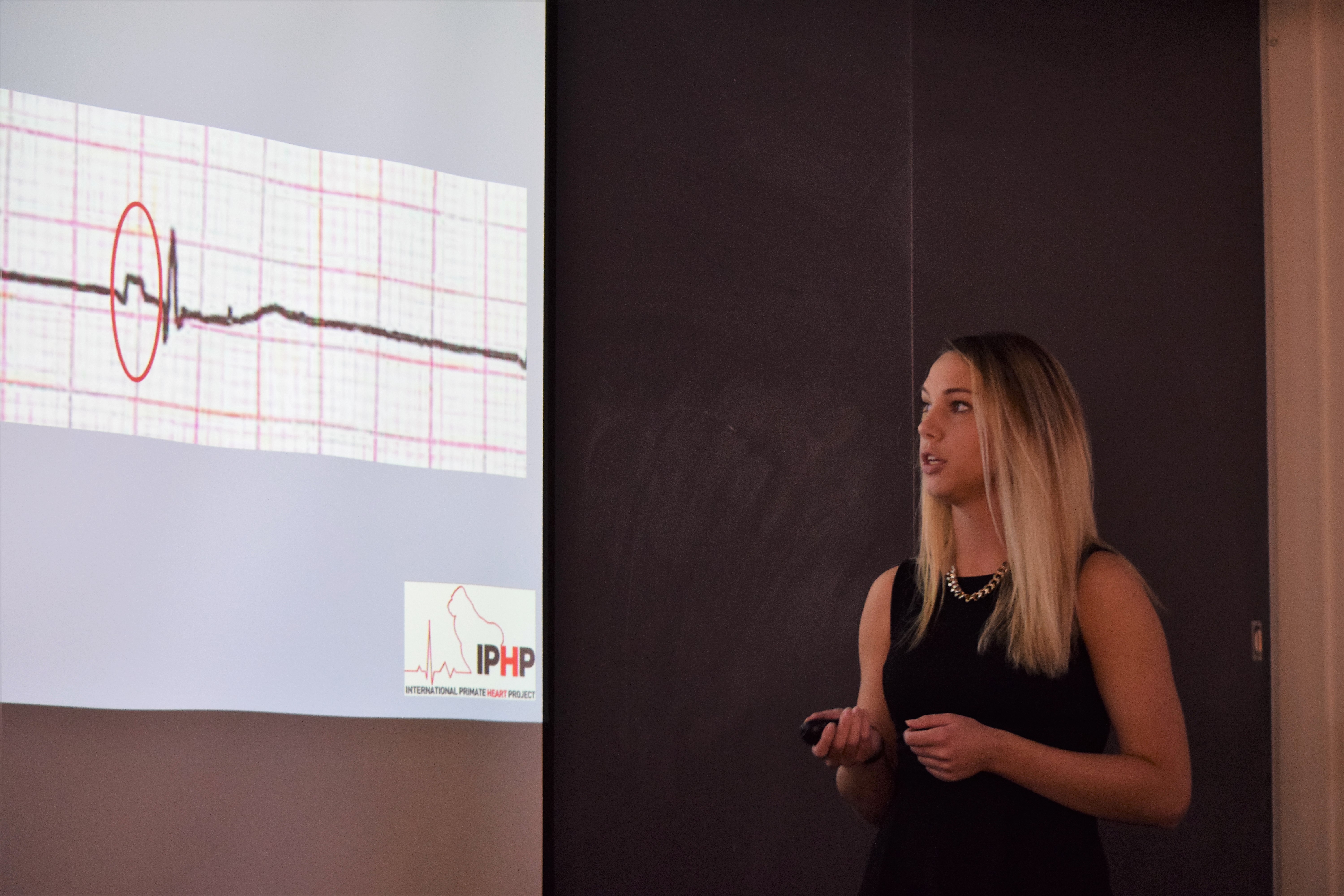
Melissa Hanson describes the results of a chimpanzee electrocardiogram at the Tropical Biology and Conservation Symposium, October 2017.
In addition to medicine, I spent much of my time analyzing behavior and social interactions of the chimpanzees. I became versed in their verbal and non-verbal language, watched alliances form, and saw individuals rise to power and dominance within their community. A chimpanzee’s well-being relies heavily on its sense of security in its social group, and the health of a chimpanzee community depends on the degree of harmony amongst its members. The knowledge base I formed through careful observation was pivotal for my participation in data collection during the integration of new chimpanzees into established social groups. During an integration, JGI caregivers and veterinarians carefully record behaviors, to ascertain whether a chimp will be accepted by its conspecifics or not. Things happen very fast, so it is imperative that observers be well-acquainted with the chimps’ social cues. In time, I was confident enough with my skills to participate in this data collection, and even had the chance to make recommendations as to which individuals to introduce to the group.
My experience did not stop there, however. While in Congo, I also had the chance to work with a variety of unique native species, such as Mandrills, a Tree Pangolin, and African Grey Parrots. During time spent with JGI’s education and public health teams, I visited local villages to discuss conservation and sustainable agriculture with their people, as well as provide parasite preventatives for their pets. I also gained a lot of experience in the laboratory analyzing blood and fecal samples and screening for infectious disease among the chimps. Additionally, I participated in several ongoing research projects at Tchimpounga and even could explore some of my own interests and questions. One endeavor I am most proud of contributing to is the establishment of a preliminary body condition score (BCS) system for chimpanzees that will allow caregivers to monitor nutrition and well-being in a non-invasive manner. Hopefully, this scoring scheme will be used when the chimpanzees are being evaluated for their success in the forest after their release.
I am incredibly fortunate to have experienced all that I did this summer, and cannot thank the partners of JGI or Engaged Cornell enough for allowing me to pursue some of my greatest aspirations while still in veterinary school. Participating in chimpanzee medicine and rehabilitation allowed me to be a part of something much bigger than myself and to learn about a species with which I had never worked, but had always dreamed of. My time in Congo made me a better student, a better person, and will undoubtedly make me a better veterinarian in the years to come.
ABOUT THE AUTHOR
 Melissa is a third-year veterinary student from Cortlandt Manor, New York. She received her Bachelor of Science degree from Duquesne University where she majored in biology and minored in biochemistry and history. Her interests are in clinical zoo and wildlife medicine and particularly rescue, rehabilitation, and release. She works as student technician at the Janet L. Swanson Wildlife Health Center, a service of the Cornell University Hospital for Animals.


