See NCBI for Complete Listing of PUblications by the rob dick Lab
Structure of the mature Rous sarcoma virus lattice reveals a role for IP6 in the formation of the capsid hexamer
Martin Obr, Clifton L. Ricana, Nadia Nikulin, Jon-Philip R. Feathers, Marco Klanschnig, Andreas Thader, Marc C. Johnson, Volker M. Vogt, Florian K. M. Schur & Robert A. Dick
Nature Communications 12 (3226), 2021.

Figure 3
a A composite model of a representative RSV CASPNC CLP, generated by placing respective unit pair averages (see groups in b) into their positions derived from subtomogram alignment. The models shown in a and in Supplementary Movie 3 are identical. b Final averages of unit pairs filtered to 8 Å. Groups are identical to their schematic depiction in Supplementary Fig. 4. Coloring of the groups corresponds with a. c Pairwise RMSD measurements between rigid-body fitted models derived from the maps of the different classes shown in b. In case of CANTD–CANTD, CANTD–CACTD, and CACTD dimer interfaces, one CA domain is fixed and RMSD is measured for the other CA domain across the respective interface. For the CACTD trimer interface one CA domain is fixed and RMSD is measured for the other two CACTD’s forming the trimer interface. d Superimposition of the models showing the flexibility of CA domains involved in the different interfaces. Color code: cyan to blue—CANTD; yellow to red—CACTD. Source data are provided with this paper.
Structures of immature EIAV Gag lattices reveal a conserved role for IP6 in lentivirus assembly
Robert A. Dick, Chaoyi Xu, Dustin R. Morado, Vladyslav Kravchuk, Clifton L. Ricana, Terri D. Lyddon, Arianna M. Broad, J. Ryan Feathers, Marc C. Johnson, Volker M. Vogt, Juan R. Perilla, John A. G. Briggs, Florian K. M. Schur

Fig 5. IP6 stabilizes the immature EIAV CASP lattice.
(A) EIAV CASP and the IP6 molecule are shown as seen from the outside of the VLP and additionally rotated by 90°. IP6 sits in the center of the hexamer and is coordinated by a ring of six lysines in the MHR (K282) and six lysines in the CASP 6HB (K351). An isosurface representation of the IP6 density is shown in pink. The densities for the individual equatorial and the axial phosphate groups are clearly visualized. The non-occupied phosphate group is caused by the 6-fold symmetry applied during processing and the fact that IP6 can sit in the binding site in 6 rotationally equivalent positions. (B) Relative infectious particle production in 293FT cells of VSV-G-pseudotyped provirus of wild type EIAV Gag (WT) and Gag with point mutations. Graphs show the average and standard deviation of three independent experiments; dots show individual data points. (C,E,G) Representative low and high magnification images of GagΔMA WT, K282A, and K351A proteins assembled in the absence (red) or presence (blue) of 10 μM IP6 at pH 6. (D,F,H) The number of VLPs (spheres-purple, tubes-green) per 55μm2 for no fewer than five representative images for each condition. Center lines show the medians; box limits indicate the 25th and 75th percentiles as determined by R software; whiskers extend to minimum and maximum values; data points are plotted as circles. The mean value of counted particles is given in italics in the bar charts.
A Structural Perspective of the Role of IP6 in Immature and Mature Retroviral Assembly
Martin Obr , Florian K. M. Schur , Robert A. Dick

Figure 1 IP6 and the retroviral life cycle.
(A) Key of the HIV-1 Gag domains on left. Right shows the d-myo-IP6 molecule. (B) 1: Gag protein interacts with the inner leaflet of the cellular plasma membrane. 2: Interactions between Gag, the plasma membrane, nucleic acid, and IP6 result in the assembly of the immature Gag lattice. 3: Assembled virion buds from cell with IP6 bound to two rings of six lysine residues at the Gag hexamer interface. 4: Retroviral protease cleaves the Gag protein, resulting in the liberation of the CA domain. CA interacts with IP6 via a ring of six arginine residues in the CANTD hexamer interface, which leads to the formation of the mature core. 5: Interaction between the viral Env protein and the cell receptor (CD4 in this example) results in fusion, and release of the viral core into the cell cytoplasm. 6: Trafficking of the viral core along microtubules to the nuclear pore. Once in the cytoplasm, dNTPs enter the capsid core where they “feed” reverse transcription of the viral RNA genome into double-stranded DNA. Once at the nuclear pore, or inside the nucleus, the capsid core breaks open, releasing the integration complex.

Figure 3 Retrovirus sequence comparison.
The immature IP6 binding site in Lentiviruses in the MHR and CACTDSPNC region (immature) and CA helix 1 (mature). Lys (K) and Arg (R) residues that are at or near known IP6 binding sites are in red. Simplified morphologies shown are on the right; conical, spherical, cylindrical, nested cores (layering). A minimum of one sequence is displayed for each retrovirus. * = retroviruses with reported IP6-related phenotypes. Italics = endogenous retroviruses. Underlined = retroviruses with solved structures with an IP6 interaction. Underlined amino acid sequences correspond to known alpha-helical structures.
Inositol phosphates are assembly co-factors for HIV-1
Robert A. Dick, Kaneil K. Zadrozny, Chaoyi Xu, Florian K. M. Schur, Terri D. Lyddon, Clifton L. Ricana, Jonathan M. Wagner, Juan R. Perilla, Barbie K. Ganser-Pornillos, Marc C. Johnson, Owen Pornillos & Volker M. Vogt

Fig. 2: IP6 interacts with Lys290 and Lys359 in the immature HIV-1 Gag hexamer.
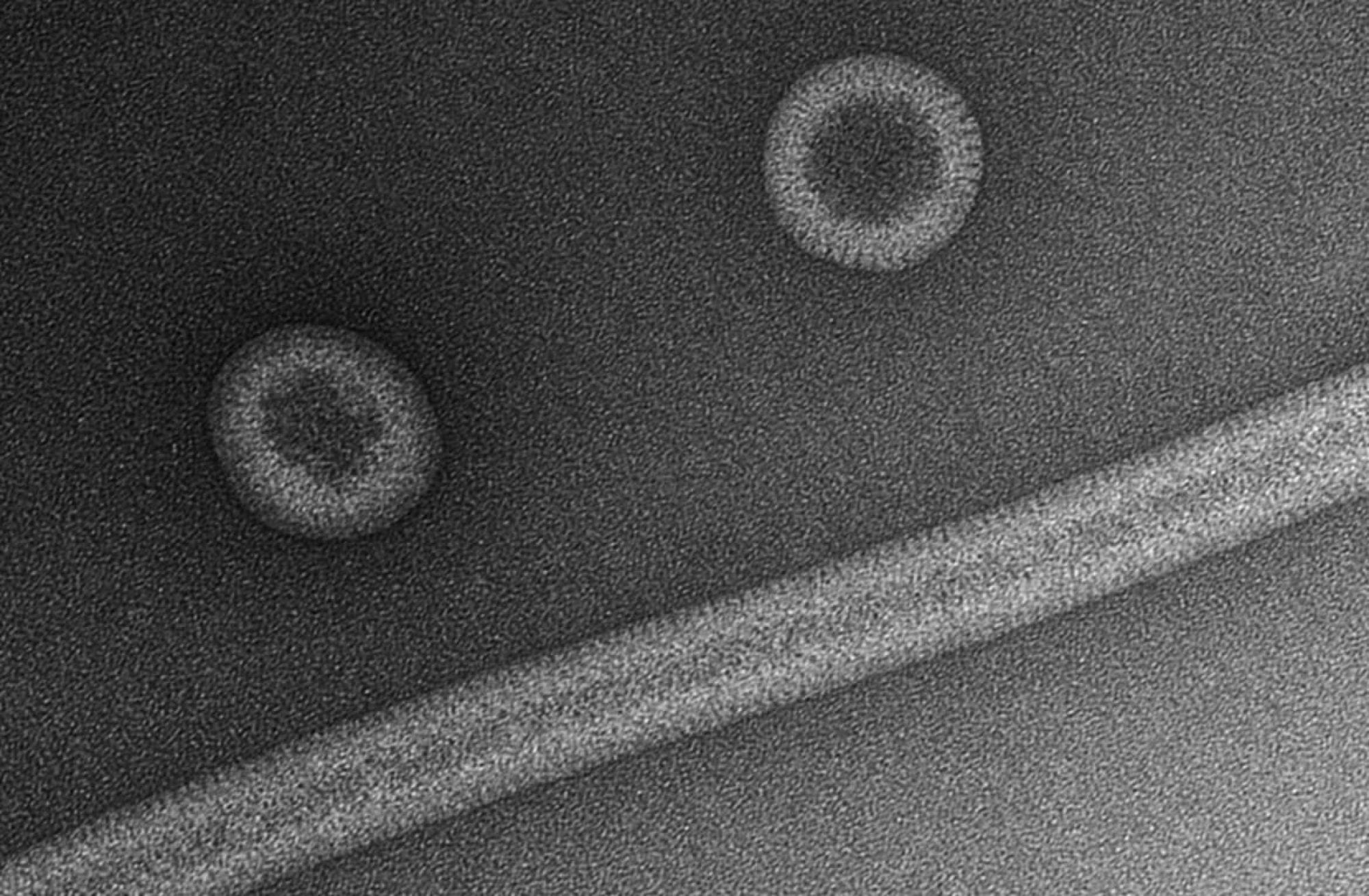
a, IP6-induced assembly of s-CASP1 into immature VLPs. The experiment was repeated four times with similar results. b, IP6-induced assembly of CACTDSP1 into flat micro-crystals. The experiment was repeated six times with similar results. c, Two-dimensional cryo-EM projection map of a micro-crystal. Images of multiple crystals were collected during two rounds of data collection from separate assembly reactions and all crystals had similar unit cells. Two individual crystals had single layer regions and could be further processed. These crystals generated similar maps. d, e, Top view (d) and side view (e) of the CACTDSP1 hexamer X-ray crystal structure showing the protein in grey ribbons and unbiased mFo–DFc difference density in blue mesh, contoured at 2σ. f, Top and side views of IP6 in its myo configuration, docked into the difference density as a rigid body in one of six rotationally equivalent orientations. All six binding modes are shown in Extended Data Fig. 4a. g, Side view of the two rings of Lys290 (green) and Lys359 (cyan) with bound IP6 in the middle. Densities were omitted for clarity, and are shown in Extended Data Fig. 4b.