Biomedical Microdevices, 2012: Immunocapture of Prostate Cancer Cells with Anti-PSMA Antibodies in Microdevices
Citation: Santana SM, Liu H, Bander NH, Gleghorn JP, Kirby BJ,
“Immunocapture of Prostate Cancer Cells with Anti-PSMA Antibodies in Microdevices”, Biomedical Microdevices, Volume 14, Number 2, Pages 401-407, 2012. doi pdf
Abstract: Patients suffering from cancer can shed tumor cells into the bloodstream, leading to one of the most important mechanisms of metastasis. As such, the capture of these cells is of great interest. Circulating tumor cells are typically extracted from circulation through positive selection with the epithelial cell-adhesion molecule (EpCAM), leading to currently unknown biases when cells are undergoing epithelial-to-mesenchymal transition. For prostate cancer, prostate-specific membrane antigen (PSMA) presents a compelling target for immunocapture, as PSMA levels increase in higher-grade cancers and metastatic disease and are specific to the prostate epithelium. This study uses monoclonal antibodies J591 and J415 antibodies that are highly specific for intact extracellular domains of PSMA on live cells—in microfluidic devices for the capture of LNCaPs, a PSMAexpressing immortalized prostate cancer cell line, over a range of concentrations and shear stresses relevant to immunocapture. Our results show that J591 outperforms J415 and a mix of the two for prostate cancer capture, and that capture performance saturates following incubation with antibody concentrations of 10 micrograms per milliliter.
Figures:
-
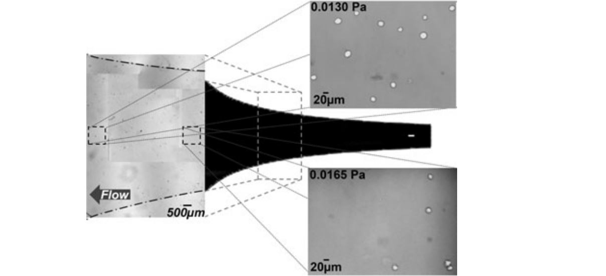
- Cell adhesion to a biotinylated-J591 immunocoated substrate at varying antibody concentrations: 10 (n09), 5 (n09), 2.5 (n09), and 1.25 μg/mL (n08), as a function of shear stress. Error bars represent the standard error of the mean; error bars are omitted from 20 microgram data for clarity
-
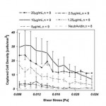
- Cell adhesion to an immunocoated substrate coated with biotinylated-J591 (n09), -J415 (n08), a 50/50 mixture of J591/J415 (n08), and NeutrAvidin (n08), as a function of local shear stress, as indicated. Error bars denote the standard error of the mean
-
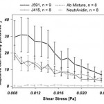
- The Hele-Shaw flow cell geometry used for these experiments is defined by the streamlines of a stagnation point flow. This form generates a linear variation in shear stress along the device’s centerline. Representative images are shown of observation fields with cells immobilized on a J591-terminated surface. The locations indicated on the image correspond to local shear stresses of 0.0165 Pa and 0.0130 Pa. The listed shear stress values correspond to a device with dimensions of: depth 48 μm, length 50 mm, inlet width 5 mm; for a volumetric flow rate of 0.2 mL/hr; with PBS. The shear stresses examined in these studies ranged from 0.008 Pa to 0.024 Pa
-
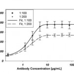
- Immunofluorescence data indicate surface coverage of immobilized biotinylated-J591 on a NeutrAvidin-coated substrate. Antibody concentrations represent the concentration of antibody in the incubating solution in micrograms per milliliter. All J591 dilutions were prepared from a stock solution of concentration 2 mg/mL. Unique curves indicate the dilution of the stock fluorophore-conjugated murine secondary antibody solution (2 mg/mL) to PBS used to stain the surface. Error bars represent standard error of the mean, all data points are representative of six repetitions (n06). Each curve was fit with a 4- parameter Langmuir adsorption isotherm. EC501:10001.5636 μg/mL, EC501:20001.3848 μg/mL