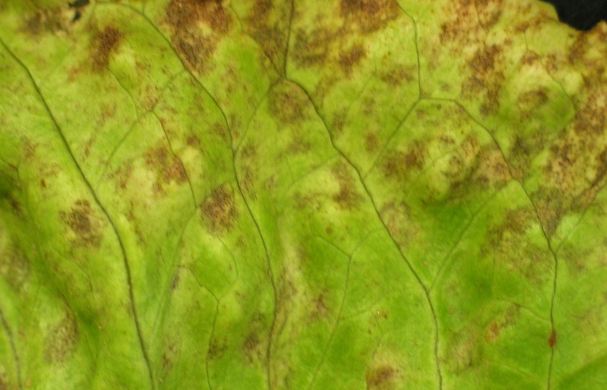
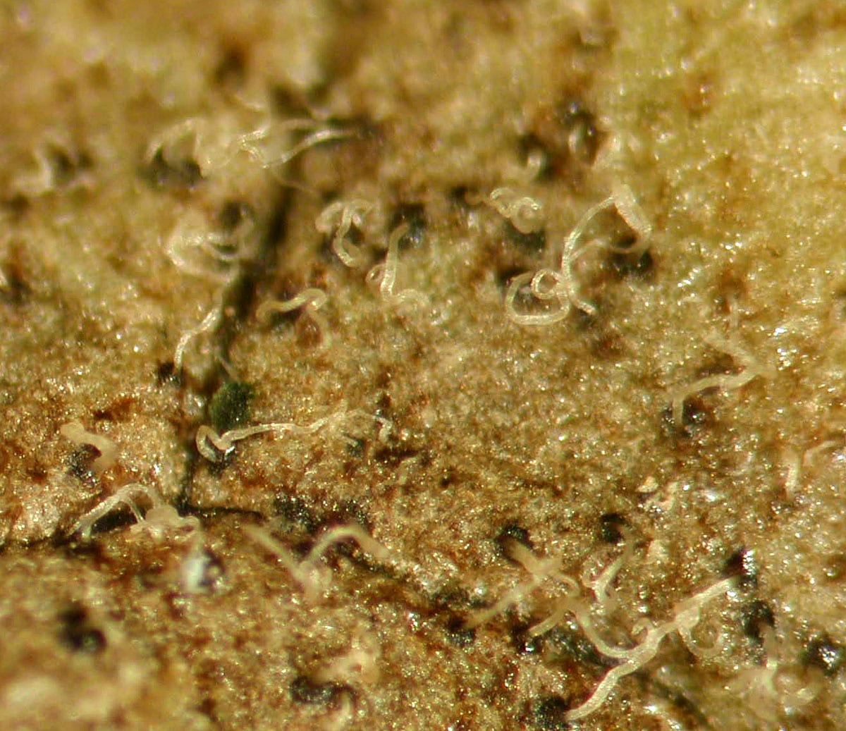
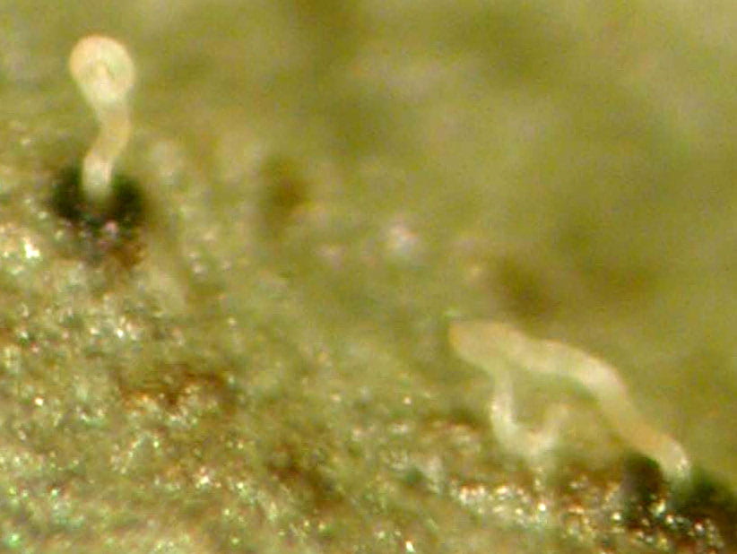
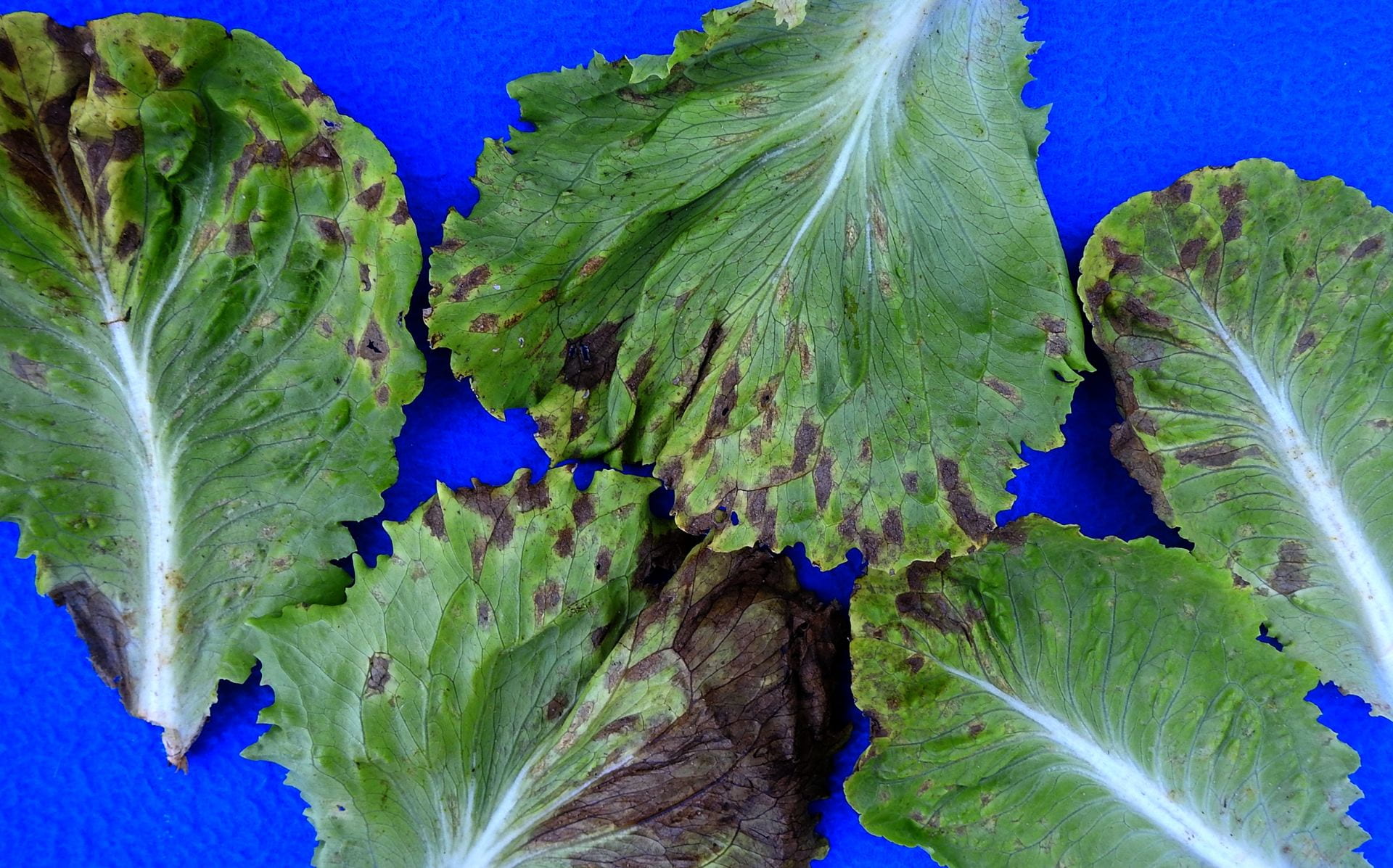
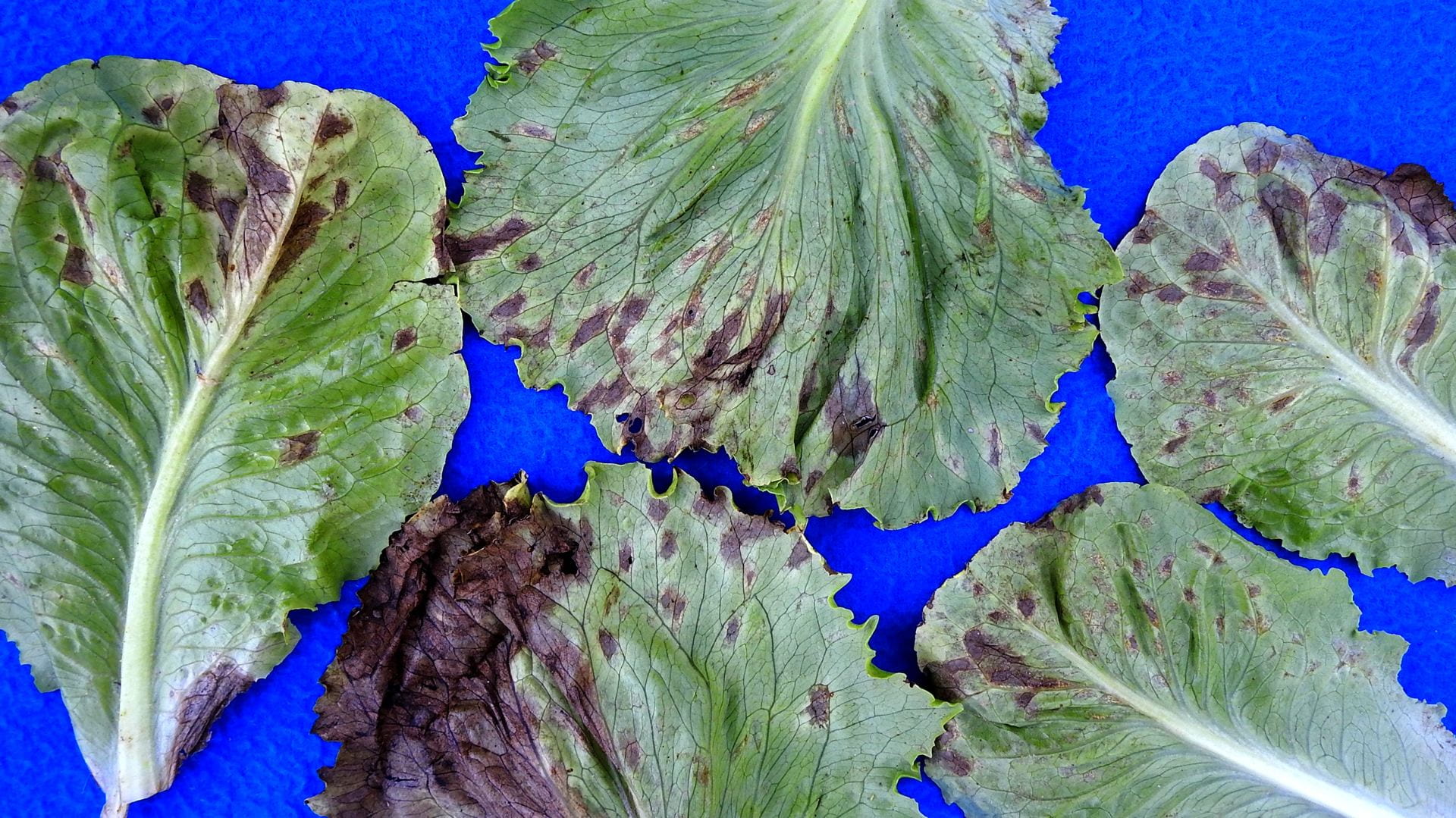
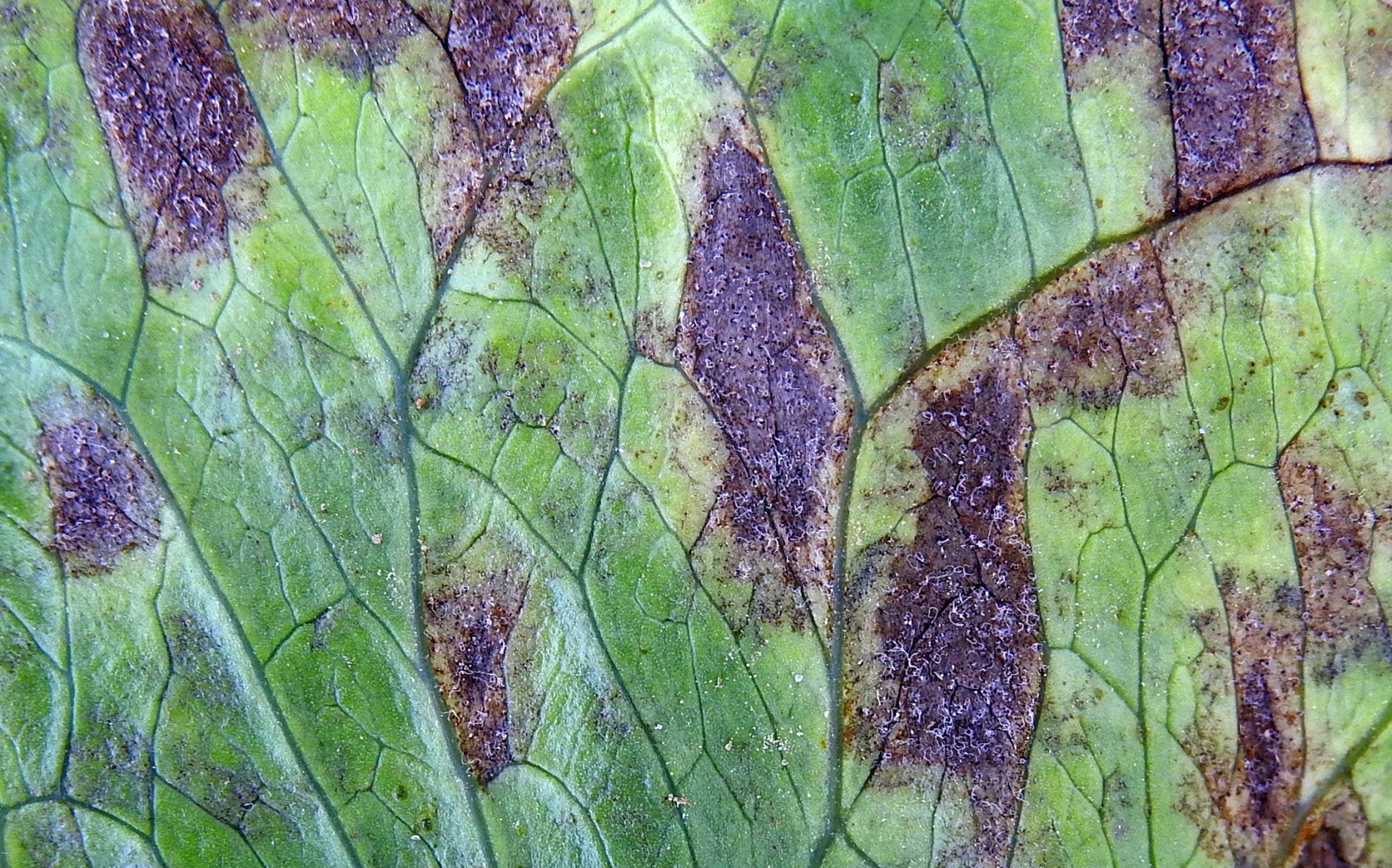
This disease has occurred rarely on Long Island with only two known occurrences from 1988 to 2022: October 2003 (first 3 images below) and June 2022 (rest). Diagnosticians in the northeast who were contacted responded they had no records or recollection of seeing Septoria leaf spot on lettuce. The pathogen produces diagnostic fungal structures (pycnidia) making it easy to identify. This disease has been reported in many countries since it was first identified in 1878, but now occurs sporadically and is considered of minor importance; however, the crop affected on LI in 2022 was unsalvageable.
The pathogen can be seed-borne. Pycnidia form on the external surface of lettuce seed. Contaminated seed is the likely source for diseases that occur rarely and are caused by pathogens like Septoria that produce spores dispersed by splashing water and thus lacking the ability to be dispersed long distances like the wind-dispersed spores of powdery and downy mildew pathogens. Hot-water seed treatment is effective but reduced germination of old seed.
Shape of leaf spots varies among lettuce varieties from circular to irregular often delineated by veins. Spots are yellow initially, then brown.
Since the pathogen is dispersed by splashing water, use drip irrigation or irrigate during the day when leaves are dry at start and will dry before nighttime to avoid long leaf wetness period.
The pathogen can survive in infested crop debris, wild lettuce, and lettuce volunteers. Thus, once introduced to a farm, it is important to remove as much infested debris as possible and/or incorporate in soil, rotate, and manage these alternate host plants.
Some information about this disease is from the Lettuce Compendium, second edition.
Below: The black dots in these lesions are pycnidia, which are diagnostic for this disease. Spore tendrils have exuded out of the pycnidia. The spores in these tendrils are dispersed by splashing water.
Following images are of upper and lower surfaces of leaves from a commercial planting on LI on 29 June 2022. Pycnidia with tendrils are visible in the close-up image of the upper leaf surface which was taken with a camera without magnification after the leaf was incubated in a plastic bag, and thus are what can be seen with limited or no magnification. Images above were taken using a dissecting microscope.


