Indonesia’s Intricacies: Rhinos, Lorises, and a Dab of Dengue

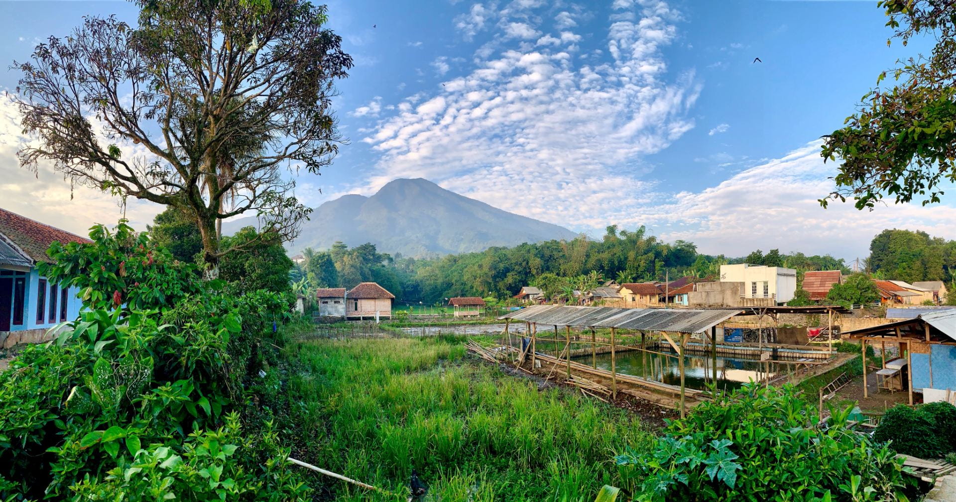
View along the way to International Animal Rescue – Bogor

When one thinks of Indonesia, picturesque scenes of Bali’s beaches or bold batik patterns might come to mind. Indonesia has an entirely different meaning to me, as this archipelago constitutes one of the most biodiverse regions on the planet. Amidst the dense rainforest foliage are fascinating endemic species, but given threats from habitat loss and poaching, the future of some Indonesian species remains uncertain. In particular, Javan and Sumatran rhinoceroses exist in a precarious state, being some of the most endangered mammals in the world. Last summer, I had the privilege of traveling to Indonesia with Carmen Smith (DVM 2021) and Montana Stone (BS 2019), under the guidance of Dr. Robin Radcliffe and support from Expanding Horizons and Engaged Cornell, to partake in a multi-faceted program centered around international conservation efforts.

Alex teaching children about the role sea birds play in the ecosystem

Building upon Dr. Radcliffe’s well-established relationships with partners in Indonesia, we were warmly welcomed by personnel from WWF-Indonesia, who had created a summer schedule with the goal of exposing us to different components of conservation work. First, we headed to the buffer zone of Ujung Kulon National Park, the last habitat of the Javan rhino. We assisted a group known as Sekala Petualang as they led conservation education programs for local school children where mainly Indonesian was spoken. It became quickly apparent that the weekly Indonesian classes we took during the spring semester before leaving were my only saving grace in that remote part of Indonesia (where cellular signals are nonexistent). Environmental stewardship was the underlying theme to the program, as educating children is an effective way to create a conservation-embracing culture. Despite living alongside a national park with Javan rhinos, a number of children were unaware that such an animal even existed in their “backyard”. It was heart-warming to see that the kids had open and receptive minds. One of Carmen’s presentations introduced veterinary medicine, yet another unfamiliarity that intrigued them. This segment of the summer underscored the importance of engaging with communities, walking in their shoes, and creating a space conducive for exchanging ideas bidirectionally. 

The next portion of the summer was spent at Institut Pertanian Bogor, a prominent university, which brought us back to the hustle and bustle of Indonesia’s urban scene. The crux of Carmen’s Expanding Horizon’s project was investigating rhino pathology as a means to better inform rhino conservation initiatives. The pathology faculty were very generous with their samples, taking us through both Javan and Sumatran rhino mortality cases. Despite my ineptitude with pathology, I was able to gain an appreciation for the challenges pathologists face working with wildlife species. I soon learned what the word “autolysis” meant as we scanned each image from the necropsy; imagine the difficulty of mobilizing a team of pathologists that must trek with their supplies to remote sites within the rainforest, finally laying their hands on the deceased rhino more than a day post-mortem, precious tissues vulnerable in the incessant Indonesian heat.

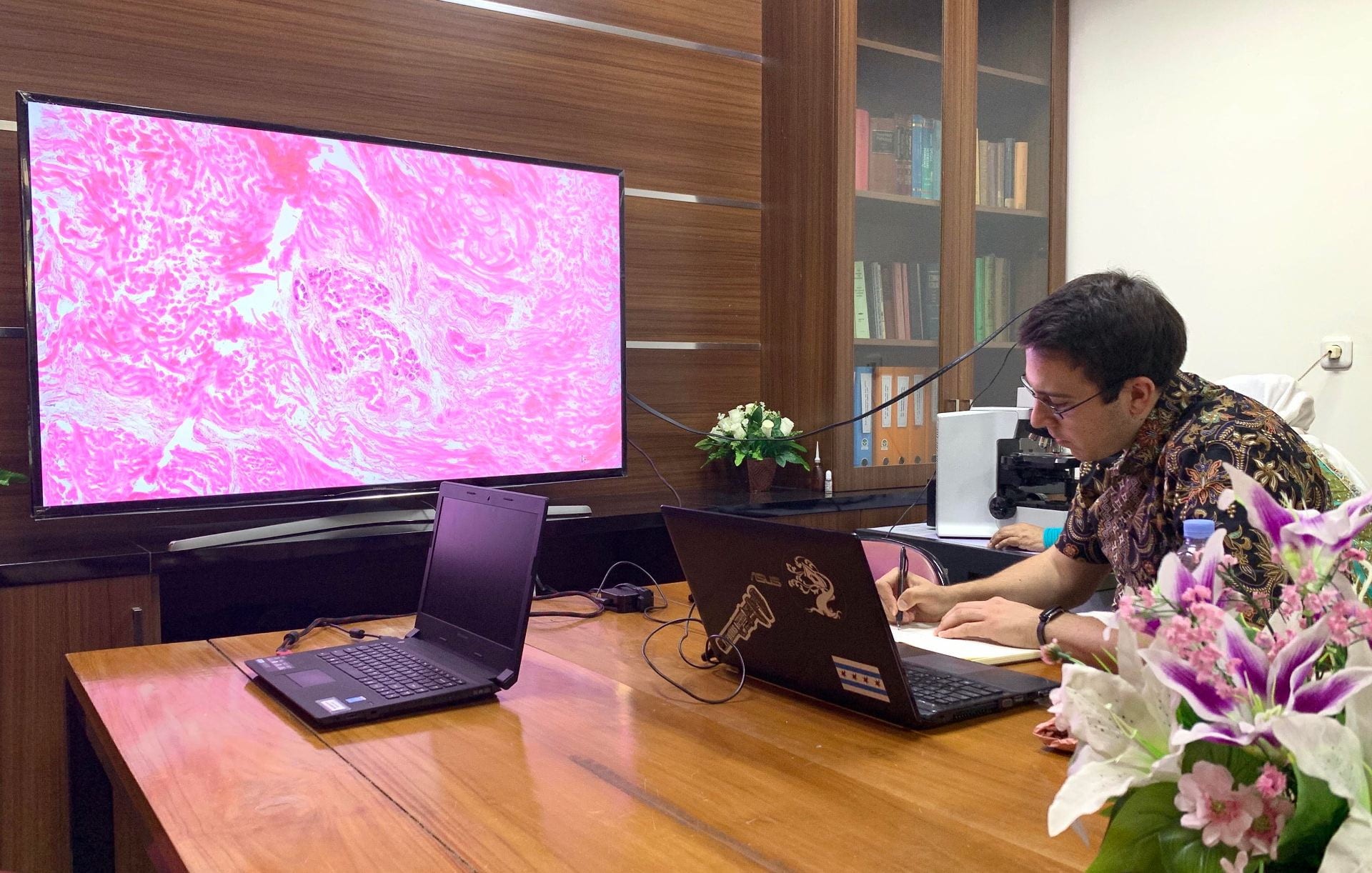
Carmen analyzing histology slides at IPB

Ultimately, Carmen and I were able to translate pathology reports into English to increase their accessibility, perform literature reviews, and identify topics worthy of discussion for a scientific publication. We realized that while individuals may have a species’ best interest in mind, other parties are bound to have conflicting interests or political underpinnings that ultimately jeopardize cohesive collaborations between various conservation organizations. We learned that any work of such nature, especially in a foreign country, must be done with exacting precision and respect. This chapter of the summer would be incomplete without mentioning my bout with dengue fever, which actually made me miss a portion of the pathology work. What started as a simple cough turned into body aches, fever, inappetence, and more. I figured an illness was inevitable in a novel tropical country, so I thought this was normal (despite running out of all of my pain medication trying to ease the symptoms). Soon thereafter, Montana also fell ill with dengue, at which point I was tested. Although no one heads off to international experiences with the intention of contracting a mosquito-borne disease, the experience was a lesson in resilience and preparedness. After all, despite being ill, I was unable to turn down a day trip to Taman Mini, a phenomenal bird park!

Prior to the next portion of the program, we briefly visited an illegal pet market, which unnerves me still to this day. I had a mental image of what such a market would entail, though nothing could have prepared me for the true horror I saw. Densely packed cages teeming with stressed birds (not to mention the dead birds littering the cage bottoms) were interrupted by cages of civets, flying foxes, macaques, and more. My heart ached for the bird that would not live to see another sunrise, for the listless civet lying motionless in its cage, and for the chained baby macaque tucked away in the shadows. I felt defeated, but I realized that such scenes are the very essence of what drives me and many other veterinary students to pursue careers that contribute to the conservation of wildlife. This experience solidified why educating the next generation on the importance of conservation and environmental stewardship is essential. 

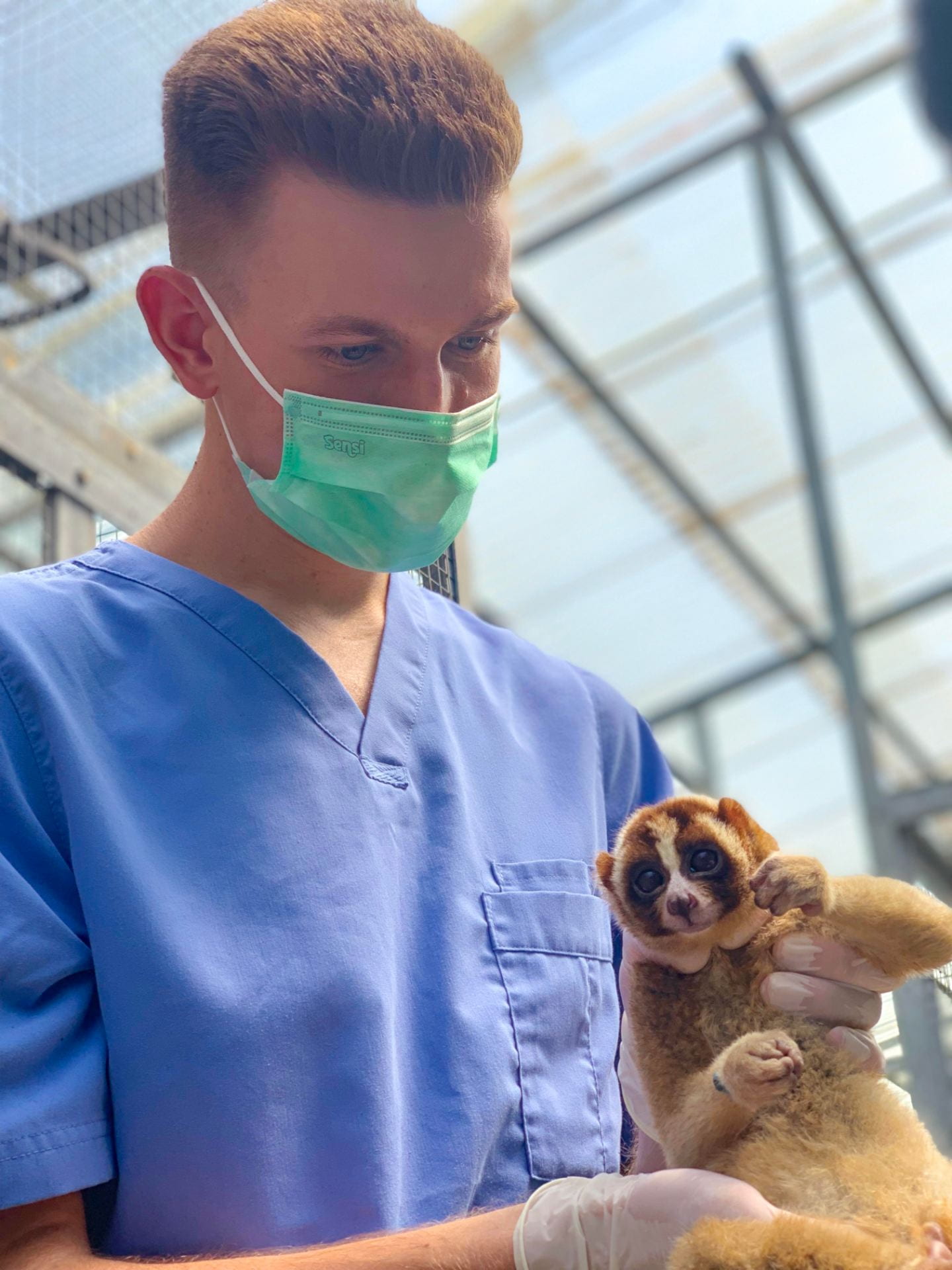
Routine health assessment of a Javan slow loris at IAR

With the atrocities from the market still fresh on our minds, Carmen and I went to International Animal Rescue – Bogor, a site that primarily focuses on rescue, rehabilitation, and release of Javan and Sumatran slow lorises. Lorises are common in the wildlife trade, and in the markets, their canines are clipped, setting the stage for dental and subsequent metabolic disease. A number of the lorises we worked with were non-releasable, so the rescue exceeded twice its anticipated capacity. We engaged in loris husbandry, spent nights observing lorises (an experience unlike any other!), and helped with their veterinary care. Multiple patients illustrated the harsh reality of a fragmented habitat. The mere existence of a road bordered by uncoated electrical wires poses a great threat to unsuspecting lorises attempting to cross the road. The prognosis is often bleak for the few lorises that manage to survive electrocution. This experience solidified my understanding of the human/wildlife dynamic, which tragically tends to swing in favor of the former.

Carmen, Montana, and Alex at Kelian Sanctuary, Borneo

Habitat loss and fragmentation set the stage for the next experience that brought us to Kelian Sanctuary in Borneo. The previous year, a Sumatran rhino named Pahu was rescued from the wild and brought to the sanctuary. Pahu is possibly only one of three rhinos remaining of her subspecies, so it is of utmost importance to rescue these isolated animals and investigate breeding options. There are Sumatran rhinos (Sumatran subspecies) that reside at the Sumatran Rhino Sanctuary in Sumatra, so collaboration is anticipated in an attempt to save this species overall. As we landed in Borneo in a precarious propellor plane, the three of us were riddled with excitement. In the few days we had at Kelian, the team composed of individuals from WWF-Indonesia and ALeRT completely immersed us in the operations at the sanctuary. Before I knew it, I was hand feeding Pahu various plants, watching her veterinary examinations and procedures, and observing her in her paddock. It was at that moment that it was clear to me that megafauna like the Sumatran rhino simply cannot be lost from this planet—it is our duty to fight with all of our passion and intensity to save such species before they slip away. Even when I was out with the team collecting food for Pahu or planting trees in the rainforest, I embraced the physical and acoustic beauty of the surrounding rainforest. During patrols of Pahu’s paddock, I witnessed gibbons, macaques, hornbills, and even a clouded leopard. During this time, Montana was able to train personnel at the sanctuary to utilize a Cornell Lab of Ornithology Swift recorder to document Pahu’s vocalizations, as no bioacoustical analysis had previously been done on Sumatran rhinos. To this day, I treasure hearing a content Pahu wallowing in the mud, “humming” and producing kazoo-like sounds, and I hope that one day everyone will know of this rhino’s plight and hear their delightful vocalizations.

As I write, I wonder whether that was my first and last time working with a Sumatran rhino. Let me re-phrase that—will the next generation even learn of critically endangered species like the Indonesian rhinos or will the species’ names be relics of a bygone era? Questions like these serve as an impetus for my passion in conservation medicine. My time in Indonesia helped me grasp what international conservation work entails, exposed me to the associated difficulties, all while leaving me inspired. I met some of the most passionate individuals in Indonesia, so I have faith that my previous questions will have favorable responses decades from now.

Diseases are Wild! A Zoo Pathology Summer Experience

Pathology is a branch of medicine related to the study of diseases, pathogens, and their effects on the afflicted animals. Pathologists can play an important role in many aspects of animal health, including detecting infectious diseases in farm animals, revealing details about animal abuse cases, and elucidating the origin cell type of a tumor from a biopsy. Some pathologists investigate diseases in wildlife species, many of which are influenced in some manner by anthropogenic factors.

For the past few summers, I have worked as a research assistant for the Zoological Pathology Program, a research and diagnostic laboratory of the University of Illinois College of Veterinary Medicine. This laboratory mostly operates in conjunction with the Chicago Zoological Society’s Brookfield Zoo, Lincoln Park Zoo, John G. Shedd Aquarium, and the Forest Preserve District of Cook County. The Zoological Pathology Program also provides diagnostics for a large number of local, national, and international conservation organizations. The faculty of pathologists in this lab are world-renowned experts in their field and serve as lead pathologists for various Species Survival Plan (SSP) programs and conservation initiatives around the globe. I am interested in the key role that pathology can play in zoo management and wildlife conservation. You can imagine, then, that I was extremely grateful to have the opportunity to work in this lab.

Throughout the three summers during which I worked in this lab, I had a wide variety of responsibilities and experiences. This work involved cataloging their extensive collection of research samples, performing molecular biology research, and observing and assisting with necropsies.

I was involved in two major research projects through the Zoological Pathology Program. One of these projects involved parasitic agents that can infect passerine, or perching, bird species. Detection of this specific infectious agent can play a particularly important part in conservation efforts involving reintroduction of captive birds, specifically the Bali Mynah. Using a diagnostic tool developed by the lab, I worked on a project aimed at determining the shedding patterns of the pathogen. The results will allow us to determine the most efficient and effective protocol for diagnostic sampling.

The other major project was a collaborative study with the Shedd Aquarium’s Microbiome Project. We investigated the efficacy of various aquarium tank disinfection techniques on the maintenance and control of atypical Mycobacteria.  Mycobacteria are common pathogens in aquatic systems and can cause fatal infections in fish. The goal of this research was to determine which disinfection techniques were better at decreasing overall loads of Mycobacterium sp. in the aquarium environment and if those disinfection techniques also altered levels of normal beneficial bacteria within these environments.  These research projects not only improve our understanding the fundamental nature of these pathogens, they also can be used as a guide to inform animal care and prevent sometimes-devastating disease outbreaks in both captive and wild populations.

In addition to those two projects, I was given the opportunity to observe and assist with necropsies of both captive and wild animals. Necropsies are the animal equivalent of forensic autopsies in humans. Although some of these necropsies (particularly those of wildlife animals) involve investigating potential criminal activity, most of the necropsies performed for zoo and wildlife animals aim to uncover infectious or management-related disease. Zoo necropsies serve to help prevent the spread of infectious disease within a zoo population, and to inform management practices to ensure optimal animal health. Information we learn about zoo animals can also inform our understanding of disease in their wild counterparts.  During my time with the Zoological Pathology Program, I participated in necropsies of various captive mammal, bird, reptile, fish, and amphibian species.  I also assisted with necropsies involving long-term seasonal assessment of disease in wild fish and reptile species in various lakes and parks. This type of study can reveal geographical patterns of disease prevalence and can potentially uncover trends associated with anthropogenic changes to the local environment.

It is our duty as animal health professionals to better understand problems to which humans contribute and to minimize their effects on wild populations. I am extremely grateful for my experiences at the Zoological Pathology Program, and I am excited to pursue a career in anatomic pathology!


ABOUT THE AUTHOR:

Carmen Smith is a current Cornell veterinary student (class of 2021) and Cornell undergraduate alumnus from the suburbs of Chicago.  He plans to pursue a career in zoo and wildlife pathology. He is interested in wildlife and public health, and hopes to work at the intersection of disease and conservation.

Lunch Lecture: “What can we really do about emerging amphibian diseases”

Wildlife Disease Association and Pathology Club are proud to present Wildlife pathologist, Dr.  María Forzán, who will speak about emerging amphibian diseases. This lecture will focus on salamander chytrid (Batrachochytrium salamandrivorans) as a potential threat to North American Species.

Thai food will be served!

Sign up for lunch here!

Please remember to bring your own plates and utensils!

The Elephant Diaries, Part 2: Field Necropsy

A 50-year-old male elephant and his loving mahout were trekking on the outskirts of a camp site when suddenly, the elephant collapsed to his death. Thankfully, this incident did not occur in front of any visitors, but the mahout was devastated nevertheless.

Given the elephant’s unexpected death, both the mahout and the camp manager wanted answers by means of a necropsy. But when the animal weighs 8,000 pounds and his body lies motionless on a dirt path in the backwoods, how do we accomplish this? Clearly, this elephant was too large and heavy to transport back to the university’s necropsy lab to perform a postmortem examination. Thus, we packed all our equipment into the mobile clinic van and traveled nearly two hours to prepare for our on-site necropsy.

When we arrived, we saw three monks circled around the elephant in prayer. Platters of food were laid painstakingly on the ground, as the monks continued their intimate ceremony in a cloud of incense. This cultural custom involves blessing the deceased elephant in order to send its spirits into the sky. During this time, the mahout gave us a medical history of his elephant, who exhibited no clinical signs prior to his death.

The process of a field necropsy is a very laborious task. Since we had no leading hypotheses about the potential cause of death, we needed to conduct an especially thorough postmortem investigation and take multiple tissue samples from various organs. The procedure would surely take us upwards of 10 hours.

Before beginning the necropsy, we first had to scout out a burial site. Fortunately, we spotted a towering tree 15 feet away. We recruited two excavator trucks to commence digging at the ground underneath. The grave needed to be several meters deep so that the elephant’s remains could be contained in a non-biohazardous manner, unlikely to get uncovered and consumed by wandering pets or wild animals. As I started putting on personal protective equipment, metal claw hands began to scrape into a ground baked hard from the Thai sun.

As the trucks continued pounding into the dirt, a tractor arrived with several motorbikes in tow, bearing chains and tarps. The tarp was laid out on the ground, where we placed our tools and organized a makeshift instrument station on its surface. At the moment, the elephant was located under an intensely hot sun, 15 feet away from the grave still being dug beside the tree. In addition, his body was currently twisted due to the way he collapsed onto the ground. Somehow, we had to both relocate him next to his shaded burial site and re-position his body into proper lateral recumbency.

That’s where the long, thick chains came into play. The chains were tied around the elephant’s limbs at one end, hurled over the tree, then connected to the rears of the motorbikes and tractor at the other end, effectively fashioning a pulley system. In unison, the tractor and motorbikes revved their engines, slowly dragging the elephant’s body inch by inch. After a few minutes of hoisting the elephant along, the tractor’s engine suddenly broke down from the heavy load, at which point they brought in a separate heavy-duty vehicle and maneuvered its crane to help nudge the elephant forward as the motorbikes continued to pull.

Once the grave was dug and the elephant was finally placed in proper position, we were ready to begin the dissection. However, all the prep work itself took roughly four hours. As storm clouds loomed in the distance, the vets and pathologists promptly took their sharpened knives and began their investigation. Plastic rope was threaded through the rim of the elephant’s skin flap so it could be reflected as they sliced through the lateral fascia. As the dissection progressed, the chains were used once again to help lift the limbs for inspection and eventual removal from the elephant carcass.

Tissue samples were handed to me to be photographed, measured, and labeled. I diligently noted gross findings as they were hollered from the vets, who were knelt down in the disemboweled abdominal cavity as they extended their hands deep into the lungs and heart.

Midway, the necropsy became a race against time when a thunderstorm descended upon us. I peered up at the tree, debating whether standing beneath it would shelter me from the downpour or electrocute me from the lightning. The camp owner’s car was parked nearby, and we rummaged through his trunk to find some rain ponchos and mini beach umbrellas. I set up shop on a plot of soil away from the tree, taking refuge under the umbrella as I waited to log in our next tissue sample.

The entire team relentlessly continued to work their way through the elephant carcass. After emerging from the body completely soaked (and not just from the rain, mind you), the vets concluded they had obtained all the necessary samples. The elephant—considerably lighter after being compartmentalized—could now be moved more easily. The crane gently goaded the carcass into the grave and scooped up the previously exhumed dirt to bury its remains. Lastly, we covered the burial site with a layer of limestone powder to help maintain the pH of the soil after the body disintegrates.

Performing an elephant field necropsy is one of the wildest veterinary procedures I have ever experienced. It combines protocol and sheer creativity, is somewhat unorthodox, and requires a concerted effort from 25 people. When resources are limited, time is tight, and logistics are threatened by weather, you can count on veterinary ingenuity and teamwork to get the job done.

Read Part 1 of The Elephant Diaries Here.

Read Part 3 of The Elephant Diaries here.

This post is written by Elvina Yau and was originally published on her WordPress blog, Elvina the Explorer, on July 26, 2017.