Biomedical Microdevices, 2014: Microfluidic isolation of cancer-cell-derived microvesicles from heterogeneous extracellular shed vesicle populations
Caption: Santana SM, Antonyak MA, Cerione RA, Kirby BJ,
“Microfluidic isolation of cancer-cell-derived microvesicles from heterogeneous extracellular shed vesicle populations “, Biomedical Microdevices, 16:869–877, 2014. doi pdf
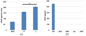
Abstract: Extracellular shed vesicles, including exosomes and microvesicles, are disseminated throughout the body and represent an important conduit of cell communication. Cancer-cell-derived microvesicles have potential as a cancer biomarker as they help shape the tumor microenvironment to promote the growth of the primary tumor and prime the metastatic niche. It is likely that, in cancer cell cultures, the two constituent extracellular shed vesicle subpopulations, observed in dynamic light scattering, represent an exosome population and a cancer-cell-specific microvesicle population and that extracellular shed vesicle size provides information about provenance and cargo. We have designed and implemented a novel microfluidic technology that separates microvesicles, as a function of diameter, from heterogeneous populations of cancer-cell-derived extracellular shed vesicles. We measured cargo carried by the microvesicle subpopulation processed through this microfluidic platform. Such analyses could enable future investigations to more accurately and reliably determine provenance, functional activity, and mechanisms of transformation in cancer.
Figures:
-
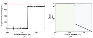
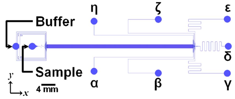
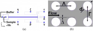
- Fig. 2 Device Performance and Transport. a Calculated Displacement. b Transport Length Ratio. doi pdf
-
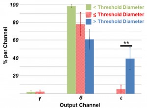
- Fig. 3 In polystyrene bead separation experiments, the microfluidic obstacle array preferentially deflects large-diameter particles in the target output. doi pdf